

- AI local brain aging maps: what USC's MRI model shows
- How AI brain age measurement works at the voxel level
- What AI local brain aging maps show in healthy adults
- Accelerated brain aging in dementia: what the ADNI data showed
- A separate genetics study points to the same vulnerable regions
- What's confirmed, and what still needs proof
- What this means for now
AI local brain aging maps: what USC's MRI model shows
USC researchers have built an AI model that estimates how old each part of the brain looks, rather than reducing the whole organ to a single age score. The team, led by Andrei Irimia of the USC Leonard Davis School of Gerontology, published the findings today in the journal Proceedings of the National Academy of Sciences, according to USC Today. The approach, an example of what researchers now call AI local brain aging maps, produces a region-by-region view of aging that lines up with brain regions known to be affected early in Alzheimer's pathology.
The model was trained on MRI scans from 14,748 cognitively healthy adults ages 19 to 100, pulled from six public datasets including the UK Biobank, the Human Connectome Project, and the Alzheimer's Disease Neuroimaging Initiative, per USC Today. Researchers then tested it on more than 1,900 additional ADNI participants: a mix of cognitively normal adults, people with mild cognitive impairment (MCI), and people with Alzheimer's disease.
The findings matter for how dementia research gets done, not for how dementia gets diagnosed. Students studying neuroscience or gerontology, caregivers following research on cognitive decline, and health-career learners tracking AI's entry into clinical neuroscience are the intended audience for what the model shows, and for the limits researchers themselves place on it.
How AI brain age measurement works at the voxel level
Brain age has been an active research area for about a decade: scientists compare a person's brain structure on MRI to patterns typical of people the same age, then flag whether the brain looks older or younger than expected, per USC Viterbi. Traditional methods typically reduce the brain to one age estimate, which can hide meaningful differences between regions, according to USC Today.
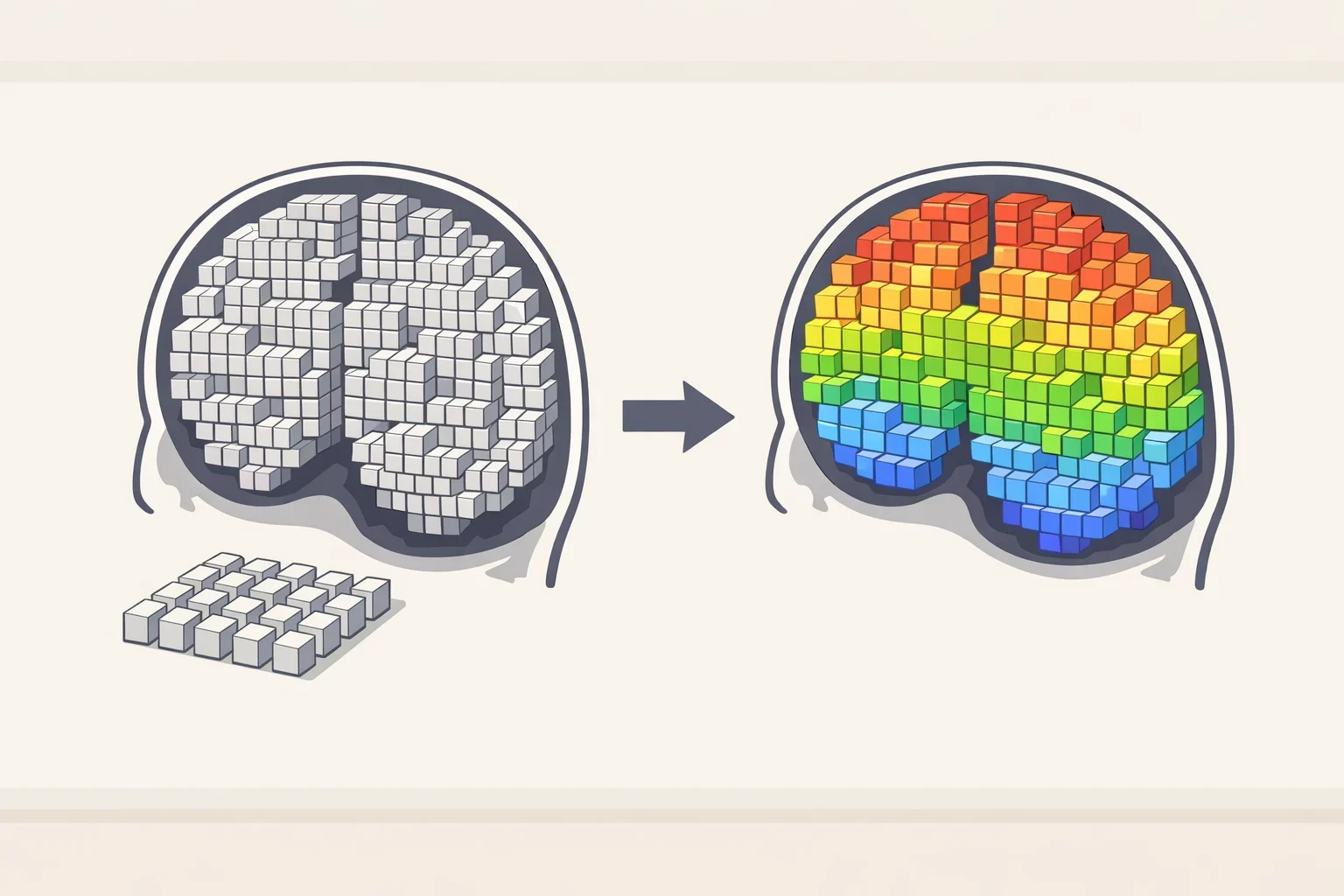
USC's model instead scores age at the voxel level, the tiny three-dimensional units that make up an MRI image, according to USC Today. A related genetics analysis using a similar imaging approach noted that a single scan contains more than two million of these units, which gives a sense of how granular this kind of mapping can get (USC Viterbi).
The output looks like a color-coded scan. Cooler colors mark regions that appear younger than a person's chronological age; warmer colors mark regions that appear older, per USC Today. In practice, a region's color reflects how it compares with the healthy reference group used to train the model, not a stand-alone medical judgment about that individual.
What AI local brain aging maps show in healthy adults

Across cognitively healthy participants, the frontal and temporal lobes, tied to decision-making and memory, consistently looked biologically older than the parietal and occipital lobes, which handle spatial awareness and sensory processing, according to USC Today. Researchers also found that the right hemisphere tended to show slightly more advanced aging than the left, a pattern that held regardless of whether a participant was right- or left-handed.
Those baseline patterns give the model a reference point for comparison. A region that looks "older" in someone with memory problems only means something in relation to that healthy baseline, and the researchers built the model around that comparison rather than around any single fixed cutoff.
Accelerated brain aging in dementia: what the ADNI data showed
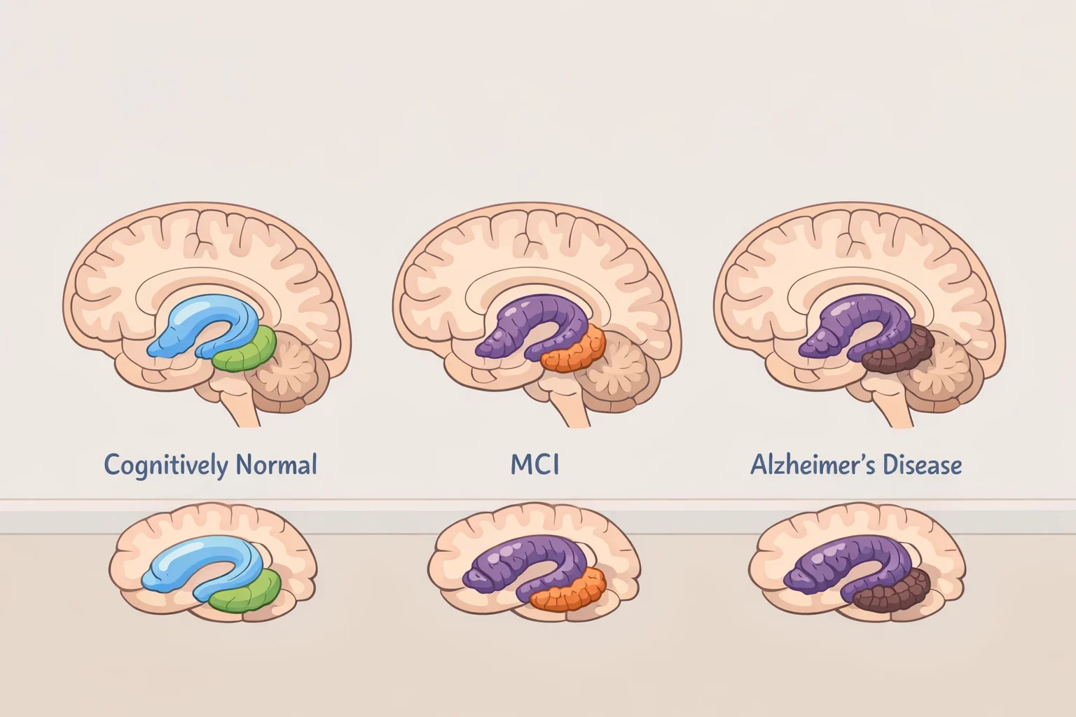
The comparison between healthy and impaired brains is where the design pays off. People with MCI or Alzheimer's disease showed significantly older local brain ages in the hippocampus, amygdala, and other structures known to be affected early in Alzheimer's pathology, per USC Today.
Compared with cognitively normal adults, impaired participants also showed more widespread advanced local aging, especially in frontal and temporal regions, and the differences grew more pronounced as impairment progressed, according to USC Today. Older local brain age also tracked with worse performance on cognitive tests, with the strongest association appearing in people already diagnosed with Alzheimer's disease.
"Not all brain regions age at the same rate," Irimia said, per USC Today. "Some areas appear to be more resilient, while others are more vulnerable to aging and disease. By measuring local brain aging, we can identify where the brain is aging faster than expected and how those changes relate to cognitive function."
Irimia described the broader stakes in similar terms elsewhere in the release. "Brain aging isn't uniform," he said, adding that tracking how individual regions age, and how those patterns differ from person to person, could eventually help identify people at risk earlier and support more personalized approaches to brain health, according to USC Today.
A separate genetics study points to the same vulnerable regions
A distinct USC-led study, published earlier this year in the journal GeroScience, adds genetic context to the pattern, though it is a separate project from the PNAS model described above. Researchers there analyzed MRI scans and genetic data from 41,708 adults in the UK Biobank, dividing the brain into 148 regions and testing more than 600,000 genetic variants against each region's aging rate, according to USC Viterbi.
That analysis turned up 1,212 significant genetic associations. One gene, KCNK2, which helps regulate electrical signaling between neurons, was linked to faster aging in regions that are especially vulnerable in Alzheimer's disease, while variants in NUAK1, involved in maintaining the structure of brain cells, were tied to younger-appearing cortex across wide areas, per USC Viterbi.
The regions that aged fastest in that genetic analysis closely matched the regions most damaged by Alzheimer's disease and frontotemporal dementia, according to USC Viterbi. Researchers there suggested accelerated local aging may be an early biological signal tied to the same vulnerability that may contribute to neurodegenerative disease later on, a hypothesis rather than a settled finding.
What's confirmed, and what still needs proof
None of this makes the USC model ready for a doctor's office. Irimia and his co-authors say the model was trained mainly on research-quality MRI and will need validation on more diverse clinical datasets before it could be used in patient care, per USC Today.
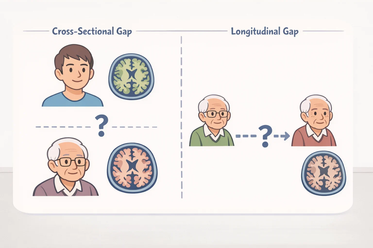
The study also relied largely on cross-sectional data, meaning it compared groups of people at a single point in time. That design cannot yet show whether local brain aging predicts which cognitively healthy adults will later develop MCI or Alzheimer's disease, a gap the researchers acknowledge directly, according to USC Today.
Other research groups are working on a related problem: making these models interpretable, not just accurate. A study published last year built a separate brain-aging tool using a technique called Shapley value interpretation to identify which specific regions drove its age predictions, then tested it on UK Biobank data. The thalamus and hippocampus turned out to be the strongest contributors to the model's predictions, and that model separated cognitively normal adults from people with MCI and Alzheimer's disease with an AUC of 0.92, according to a bioengineering study. That's a different tool built on a different dataset, so it doesn't validate USC's model, but it points in the same direction: regional aging patterns that track meaningfully with disease status.
The caution extends across the wider field of AI dementia tools. A review published last year of 21 studies covering more than 1 million participants found these models perform well on average, with a mean AUC of 0.845, but noted that external validation remained limited and that calibration and generalizability were ongoing problems, according to an Europe PMC systematic review.
What this means for now
Taken together, the findings describe a research tool that lines up with regions known to be damaged early in Alzheimer's disease. A separate genetics study and a separate interpretability-focused aging model offer related, preliminary evidence from different approaches, but neither one validates USC's model for predicting an individual's future decline. Irimia's team describes the model as useful for studying disease mechanisms and potentially tracking whether experimental treatments slow degeneration in specific regions, not for diagnosing any one patient, according to USC Today.
Anyone following this research for a class project, a caregiving decision, or personal curiosity should read the peer-reviewed PNAS paper and watch for future longitudinal validation studies rather than treating a colorful brain map as a diagnosis. People with real concerns about memory or thinking changes, their own or a family member's, should raise them with a primary care doctor or neurologist who can order the appropriate clinical evaluation. A research-stage MRI model is not a substitute for that conversation.